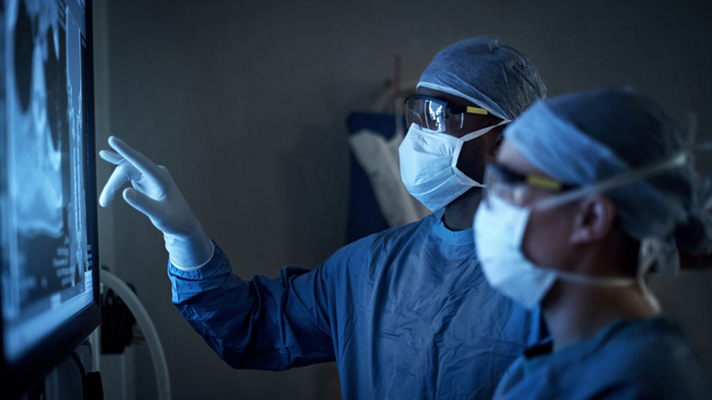
"If you have a radiologist sitting and doing regular bread and butter imaging, AI can help in accelerating those imaging exams.”
That’s according to Siddharth Shah, healthcare industry analyst at Frost & Sullivan, who in a recent commentary suggested the innovation phase for AI in radiology is nearing its end, as healthcare organizations have started adopting AI tools for radiologists and the tools have proved useful.
For example, as tech writer Makenzie Holland explains, at University of Utah Health, Richard H. Wiggins III, M.D., is using a new tool to seamlessly compile a patient's prior scans.
Wiggins, who is director of imaging informatics and medical administrator for PACS at the University of Utah Health, spends a lot of time each day looking at hundreds of medical images. He often needs to see not only a patient's current CT scan or other medical image, but also prior scans to compare how an abnormality is developing.
In the “old days,” Wiggins had to perform a manual search of the archive and then assemble the images together in his workflow to view them. The process was time-consuming and inefficient, so Wiggins went looking for a way to make prior scans easily accessible.
Now, he is able to draw information from data sources across his healthcare organization, such as the EHR, for patient analysis, and then pull images into the radiologist's workflow. Wiggins can then interact with the scans and even click on specific features to pull up related examples.
At the recent Radiological Society of North America's annual meeting, Wiggins said that with the right-click of a mouse, he can pull older scans from the archives, arrange them according to relevance and have them right in front of him for reference.
"It's difficult sometimes to search for old studies, so the ability to right mouse click on something and say 'find all the prior post-contrast imaging and put them next to each other so I can quickly compare these,' that's very valuable for my time," Wiggins said.
Another example Holland cites is that of Pennington, NJ-based Capital Health Hospitals' Ajay Choudhri, M.D., who is using an AI-based clinical tool to identify brain bleeds in CT scans.
Choudhri, chairman and medical director of the department of radiology at Capital Health Hospitals in New Jersey, has been using AI to detect brain bleeds in CT scans, which he said has helped his department better prioritize critical cases.
He uses AI-enabled software to quickly identify intracranial hemorrhages in CT scans, and then prioritize the scans within radiologists' work lists.
"There are a lot of scans that are negative that can move down the list so we can attend to people who are more sickly, instead of it sitting on the list waiting to be read," Choudhri said.
The ability to flag and prioritize a patient with a suspected brain bleed can save time and expedite patient care, particularly for healthcare organizations that might not have a neuroradiologist on hand, he said.


